


- HOME
- New Product and Technology
- Protease Inhibitor
Protease Inhibitor 2026/08/25
Protease inhibitors are essential for minimizing degradation of target proteins during protein purification from plant and animal cells or tissues. When tissue is homogenized (disrupted), proteases that were previously sequestered within cellular organelles, particularly lysosomes, are released into the lysate. Once these proteases come into contact with the target protein, degradation can proceed rapidly. Performing the entire procedure at a low temperature (4 °C) can reduce proteolysis to some extent; however, appropriate protease inhibitors are indispensable when tighter control is required.
1. Protease Classes and Criteria for Selecting Inhibitors
To control proteases released during cell disruption, inhibitors should be selected on the basis of two factors: the catalytic mechanism (protease class) and the selectivity of each inhibitor for individual enzymes.
Selection Based on Catalytic Mechanism (Protease Class)
Proteases are classified mainly into the following four groups according to the structure of their active site and their catalytic mechanism.
- Serine proteases (e.g., trypsin-like and chymotrypsin-like proteases, plasmin, and kallikrein)
- Cysteine proteases (e.g., papain-like proteases and cathepsin B)
- Aspartic proteases (e.g., pepsin-like proteases and cathepsin D)
- Metalloproteases (e.g., thermolysin and carboxypeptidase A)
Small-molecule inhibitors generally act at or near the active site and block protease-catalyzed hydrolysis. A practical first step is therefore to select an inhibitor that matches the catalytic class of the protease expected in the sample.
Selective Inhibition of the Target Protease
Even within the same protease class, inhibitor selection must take substrate specificity into account when a particular enzyme needs to be inhibited selectively. Among inhibitors of serine and cysteine proteases, for example, chymostatin preferentially inhibits chymotrypsin-like proteases, whereas leupeptin and antipain do not show comparable inhibition of α-chymotrypsin. Understanding the characteristic inhibition profile of each compound is therefore important.
Building an Inhibitor Cocktail for the Experimental System
As described above, proteases differ in both catalytic mechanism and substrate specificity. To suppress the range of proteolytic activities that may coexist in a lysate, inhibitors with complementary mechanisms are commonly combined in a protease inhibitor cocktail.
Commercial premixed cocktails are convenient, but their composition can be difficult to fine-tune for a particular experimental system. Peptide Institute supplies individual protease inhibitors, allowing researchers to build and adjust a cocktail according to their experimental design.
Typical Situations in Which Cocktail Optimization Is Required
- When the target protein is itself a protease or another enzyme whose activity may be affected
If the protein being recovered and purified is a protease, a premixed cocktail may also inhibit the desired target activity. Selecting only the necessary components makes it possible to preserve the activity of the target enzyme while suppressing degradation caused by unwanted proteases. - When an inhibitor may interfere with downstream analysis or experiments
Certain inhibitors, including irreversible cysteine-protease inhibitors such as E-64, may affect subsequent mass spectrometry (MS) or cell-based assays. Excluding only the components that are incompatible with the downstream workflow can help minimize such interference.
2. Detailed Profiles of Individual Protease Inhibitors
| Inhibitor | Primary Target Proteases | Typical Working Concentration | Solvent | Inhibition Mode | Evidence / Notes |
|---|---|---|---|---|---|
| Chymostatin | Serine (chymotrypsin-like) and selected cysteine proteases | 10–100 µM | DMSO | Reversible | Structure and properties reported[1]; slow-binding inhibition reported[2] |
| Leupeptin | Serine and broad-range cysteine proteases | 10–100 µM | H2O | Reversible | Inhibits plasmin, trypsin, and papain, but not α-chymotrypsin[3] |
| Antipain | Serine and cysteine proteases (complementary coverage) | 1–10 µM | H2O (1 mM) | Reversible | Reported as a novel protease inhibitor[4] |
| E-64 | Cysteine proteases (primary choice) | 10 µM | H2O | Irreversible | Properties reported for the novel inhibitor[5]; inhibition mechanism discussed[6] |
| Pepstatin A | Aspartic proteases | 1 µM | DMSO or EtOH:AcOH 9:1 (1 mM) | Reversible | Reported as a novel pepsin inhibitor[7]; inhibition mechanism reported[8] |
| Aprotinin | Serine proteases | 2–10 µg/mL | H2O | Reversible | Reviewed as a serine-protease inhibitor[9] |
2.1 Chymostatin
Chymostatin is a peptidic inhibitor that was identified and structurally characterized as a chymotrypsin inhibitor. Kinetic analysis has demonstrated potent slow-binding inhibition of chymotrypsin and cathepsin G[1,2].
Structure and mode of action: The C-terminal aldehyde group forms a reversible hemiacetal intermediate with the active-site serine residue of the target enzyme, or a hemithioacetal with an active-site cysteine residue, thereby inhibiting catalysis. Formation of the final high-affinity complex is time dependent, giving rise to slow-binding behavior.
2.2 Leupeptin
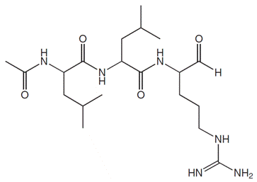
Leupeptin strongly inhibits plasmin, trypsin, and papain but does not inhibit α-chymotrypsin, demonstrating a distinct substrate-specificity profile[3].
Structure and mode of action: Inhibition is mediated by the C-terminal aldehyde group. The arginine residue at the P1 position contributes to selectivity for trypsin-like proteases.
2.3 Antipain
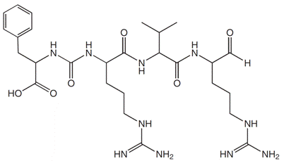
Antipain is an actinomycete-derived protease inhibitor that has long been used as a component of custom inhibitor cocktails. Trypsin is among its principal targets[4].
Structure and mode of action: Inhibition is mediated by the C-terminal aldehyde group.
2.4 E-64
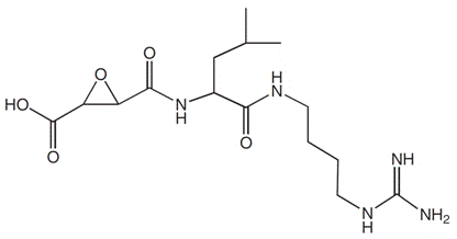
E-64 was isolated as a novel thiol-protease inhibitor and is a potent inhibitor that binds irreversibly to papain, cathepsin B, and related enzymes at an equimolar ratio. Structure–activity studies of enzyme–inhibitor complexes have clarified its detailed mechanism of action. Derivatives with improved cell permeability, including E-64-d, have also been developed[5,6].
Structure and mode of action: The epoxysuccinyl moiety (epoxide ring) undergoes a selective nucleophilic reaction with the active-site thiol group of cysteine proteases. This irreversibly S-alkylates the catalytic cysteine and inhibits enzymatic activity.
2.5 Pepstatin A
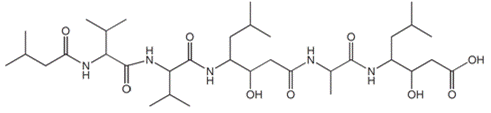
Pepstatin A was discovered as an actinomycete-derived pepsin inhibitor and is widely used as a representative inhibitor of aspartic proteases. Pepsin is among the major targets for which inhibitory activity has been reported[7,8].
Structure and mode of action: Pepstatin A contains statine, an unusual non-proteinogenic amino-acid residue. This moiety mimics the transition state of protease-catalyzed hydrolysis. By binding noncovalently within the active-site pocket of an aspartic protease as a transition-state analogue, pepstatin A inhibits catalysis.
2.6 Aprotinin
Aprotinin inhibits a broad range of serine proteases, including trypsin, chymotrypsin, plasmin, and kallikrein, and is widely used as a general-purpose serine-protease inhibitor[9].
Structure and mode of action: Aprotinin is a proteinaceous inhibitor consisting of 58 amino-acid residues. Unlike small-molecule inhibitors that react through functional groups such as aldehydes or epoxides, aprotinin binds tightly in the target enzyme’s active-site cleft in a substrate-like manner without relying on such reactive groups.
3. Essential Experimental Practices for Minimizing Proteolysis
Recovering the target protein at high yield requires not only appropriate inhibitor selection but also careful sample handling. The following four practices help minimize protease activation and improve recovery of the target protein[10].
- Keep the sample cold (4 °C)
To minimize intrinsic protease activity, perform all operations at 4 °C or on ice. Prechill buffers and equipment, and keep the sample on ice until immediately before centrifugation. - Add inhibitors immediately before homogenization
Some inhibitors lose activity depending on buffer pH, composition, or elapsed time. Add them to the prechilled buffer immediately before disruption so that an effective concentration is present during homogenization. Adding inhibitors only after tissue disruption should be avoided because degradation may begin within the short interval immediately after lysis. - Minimize frictional heating by keeping processing times short
Mechanical friction generated by a blender or homogenizer can produce local heating, leading to thermal denaturation of the target protein or unintended activation of proteases. Limit processing to the shortest practical duration and use intermittent operation with cooling. - Mix promptly after homogenization to eliminate local concentration gradients
Immediately after cell or tissue disruption, localized regions containing high concentrations of proteases may arise within the lysate. Mix the entire sample promptly to homogenize it and distribute the inhibitors throughout the solution.
Product List
| Code | Product | Package |
|---|---|---|
| 4063 | Chymostatin | 25 mg / 100 mg |
| 4041-v | Leupeptin | 0.5 mg |
| 4041 | Leupeptin | 25 mg / 100 mg / 1 g |
| 4062-v | Antipain | 0.5 mg |
| 4062 | Antipain | 25 mg / 100 mg |
| 4096-v | E-64 | 0.5 mg |
| 4096 | E-64 | 25 mg / 100 mg / 1 g |
| 4397-v | Pepstatin A | 0.5 mg |
| 4397 | Pepstatin A | 25 mg / 100 mg / 1 g |
| PRO-285 | Aprotinin | 100 mg / 250 mg / 1 g |
References
- K. Tatsuta et al., “The structure of chymostatin, a chymotrypsin inhibitor.” J. Antibiot. (Tokyo), 26(11), 625–646 (1973). DOI: 10.7164/antibiotics.26.625.
- R. L. Stein and A. M. Strimpler, “Slow-binding inhibition of chymotrypsin and cathepsin G by the peptide aldehyde chymostatin.” Biochemistry, 26(9), 2611–2615 (1987). DOI: 10.1021/bi00383a030.
- T. Aoyagi et al., “Biological activities of leupeptins.” J. Antibiot. (Tokyo), 22(11), 558–568 (1969). DOI: 10.7164/antibiotics.22.558.
- H. Suda et al., “Antipain, a new protease inhibitor isolated from actinomycetes.” J. Antibiot. (Tokyo), 25(4), 263–266 (1972). DOI: 10.7164/antibiotics.25.263.
- K. Hanada et al., “Isolation and characterization of E-64, a new thiol protease inhibitor.” Agric. Biol. Chem., 42(3), 523–528 (1978). DOI: 10.1271/bbb1961.42.523.
- K. Matsumoto et al., “Structural basis of inhibition of cysteine proteases by E-64 and its derivatives.” Biopolymers, 51(1), 99–107 (1999). DOI: 10.1002/(SICI)1097-0282(1999)51:1<99::AID-BIP11>3.0.CO;2-R.
- H. Umezawa et al., “Pepstatin, a new pepsin inhibitor produced by actinomycetes.” J. Antibiot. (Tokyo), 23(5), 259–262 (1970). DOI: 10.7164/antibiotics.23.259.
- J. Marciniszyn Jr., J. A. Hartsuck, and J. Tang, “Pepstatin inhibition mechanism.” Adv. Exp. Med. Biol., 95, 199–210 (1977). DOI: 10.1007/978-1-4757-0719-9_12.
- A. V. Ivachtchenko et al., “Aprotinin—Drug against Respiratory Diseases.” Int. J. Mol. Sci., 24(13), 11173 (2023). DOI: 10.3390/ijms241311173.
- S. Roe (ed.), Protein Purification Applications: A Practical Approach, 2nd ed., Oxford University Press (2001). Academic methods volume containing examples of protease-inhibitor cocktail implementation. DOI: 10.1093/oso/9780199636723.001.0001.